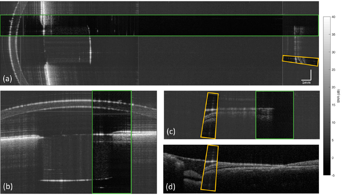
A team of researchers has developed a new optical scanner system that allows high-quality images of the entire eye to be obtained economically and without the need for expensive mechanical components. This innovative technology, published in the journal Biomedical Optics Express, has the potential to increase the accessibility of diagnosis and surgical planning oculars, as well as expanding its use in various ophthalmic applications.

The researchers carried out experimental tests to validate the effectiveness of the optical scanner. Using an artificial human eye model and an ex vivo rabbit eye, they acquired OCT images of the entire eye and compared the results with those obtained using conventional OCT systems. The results showed comparable characteristics and satisfactory image quality, demonstrating the clinical potential of this new technology.
The new optical scanner offers numerous advantages in the field of ocular biometry, cataract surgery planning, glaucoma diagnosis and severe myopia progression studies. Furthermore, its dynamic reconfiguration capacity opens the door to new ophthalmic applications and future developments.
This innovation represents a significant advancement in the field of ophthalmology by offering a versatile, cost-effective and high-quality solution for whole-eye imaging. This technology is expected to have a positive impact on ocular healthcare and contribute to early and accurate diagnosis of various ocular diseases.
This research work is a collaboration between the spin-off 2EyesVision S.L., the CSIC Institute of Optics, the International Center for Translational Eye Research (ICTER) of the Institute of Physical Chemistry of the Polish Academy of Sciences (IPC-PAS) and the Center for Visual Science of the University of Rochester from NY
IO-CSIC Communication
cultura.io@io.cfmac.csic.es
Related news
New in the lab: Elena Moreno and Diego Dijkstra
Elena Moreno Rubio, BSc in Physics by the University of Murcia and current MSc student in Optical and Imaging Technologies at the Complutense...
Diagnosis of ocular diseases by ocular biomechanics
Tomorrow, January 25, VioBio Lab researcher Judith Birkenfeld will give a guest lecture at the Fernández Vega Foundation as part of the Fernández...
SureVision, one of the 10 projects selected by IMPULSA-T 2023
The SureVision project lead by our colleague, Víctor Rodríguez López, has been one of the 10 projects selected by the Impulsa-T program, focused on...


